Key benefits
Multiscale super-resolution
By combining high-throughput spinning-disk confocal with high-resolution SIM, researchers can unlock the full potential of multi-scale imaging.
Unparalleled versatility
The synergistic technology combination offers users the freedom to image a wide range of sample types.
Multimodal super-resolution
SIM combined with SPIN SACD super-resolution offers dual super-resolution technologies on a single platform.
Multiscale super-resolution
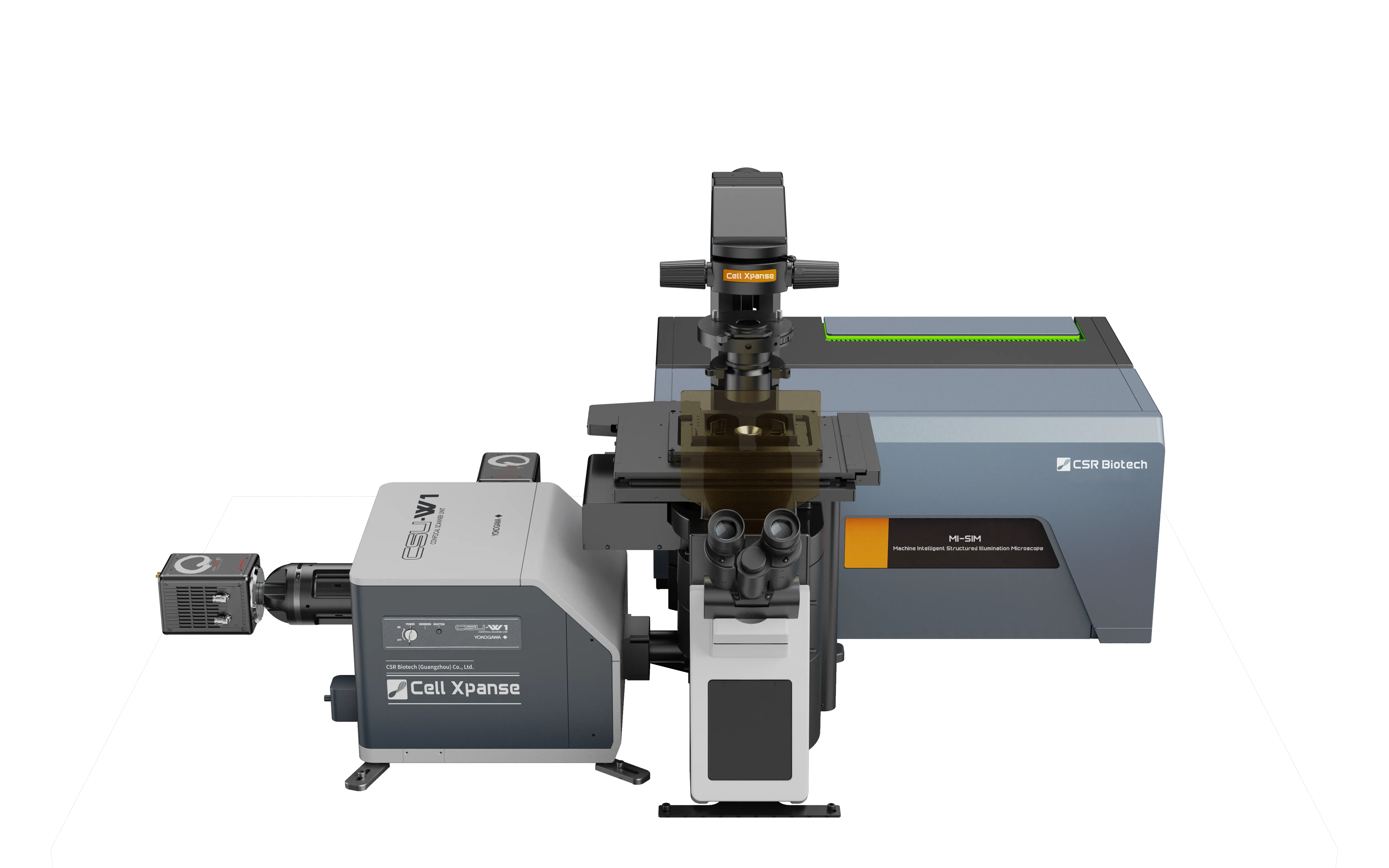
Spinning disk offers a high-throughput confocal imaging solution that can rapidly scan large format samples. When combined with high resolution capabilities offered by SIM, the Cell Xpanse system enables users to explore samples across multiple resolution scales.
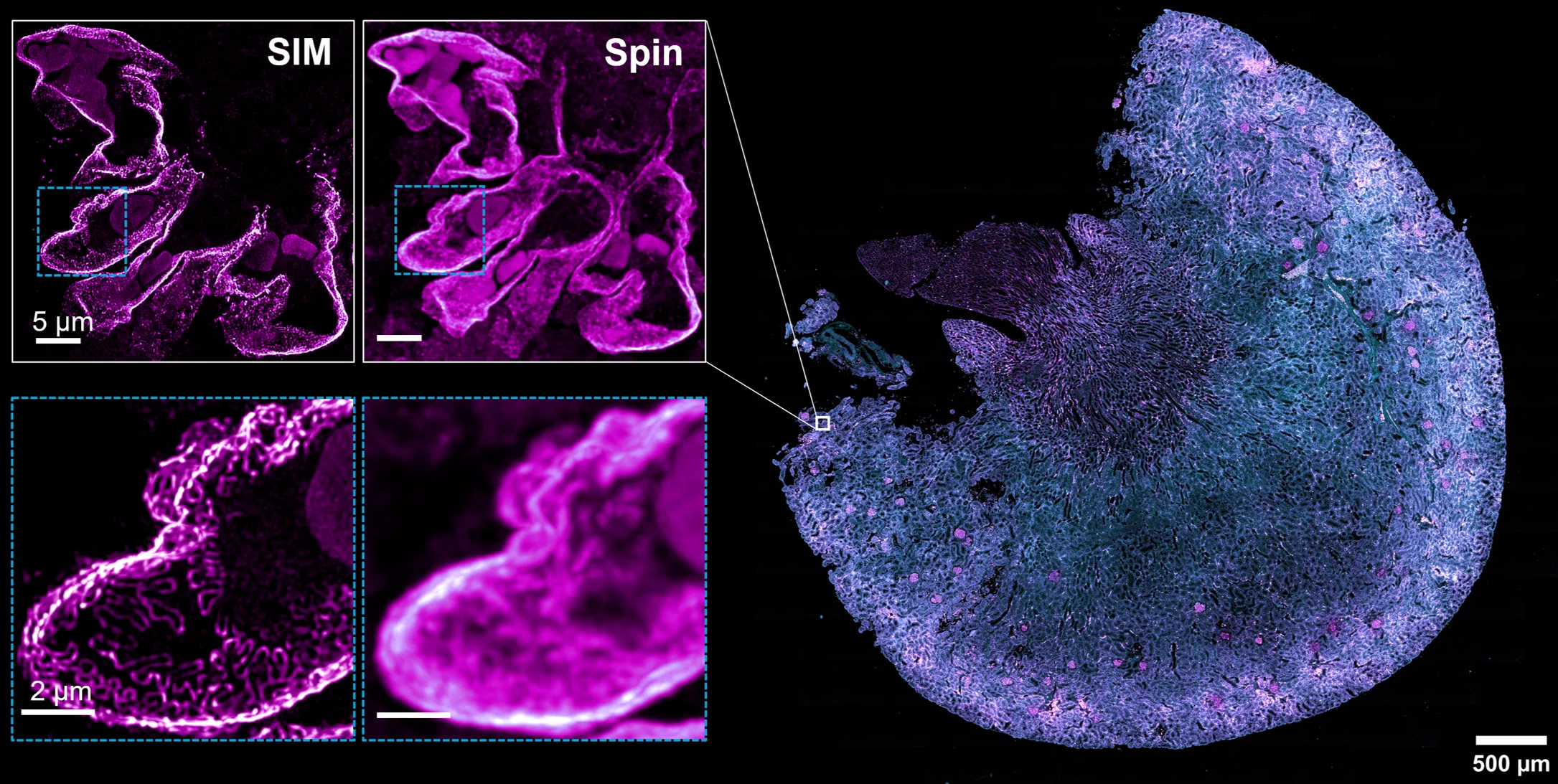
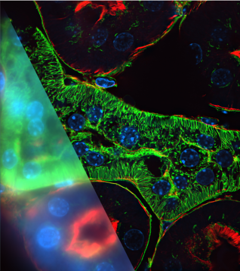
Figure shows a kidney section, courtesy of Nipoka. The sample was imaged with spinning disk. Zoomed regions reveal that spinning disk cannot fully resolve podocytes. Once a region of interest is identified, SIM can be used to resolve the podocyte structure.
Advanced workflow and system integration
Imager software enables users to combine multi-imaging modalities, multi-site imaging and multi-phase imaging into a single workflow.
Advanced deconvolution and AI denoising tools can be used to remove residual pinhole crosstalk from spinning-disk data, revealing detailed 3D images.
Together with 3D visualization and batch processing tools, users can process complex biological datasets into high-quality results.
This all-in-one solution empowers researchers to streamline their workflows and unlock new insights from their complex biological data sets.
Figure shows Imager software set up to image 20 separate regions. Event-based AI triggers monitor for mitosis. Upon detection of a mitotic event, the system activates a second channel and reduces the time-lapse interval.
Multimodal super-resolution
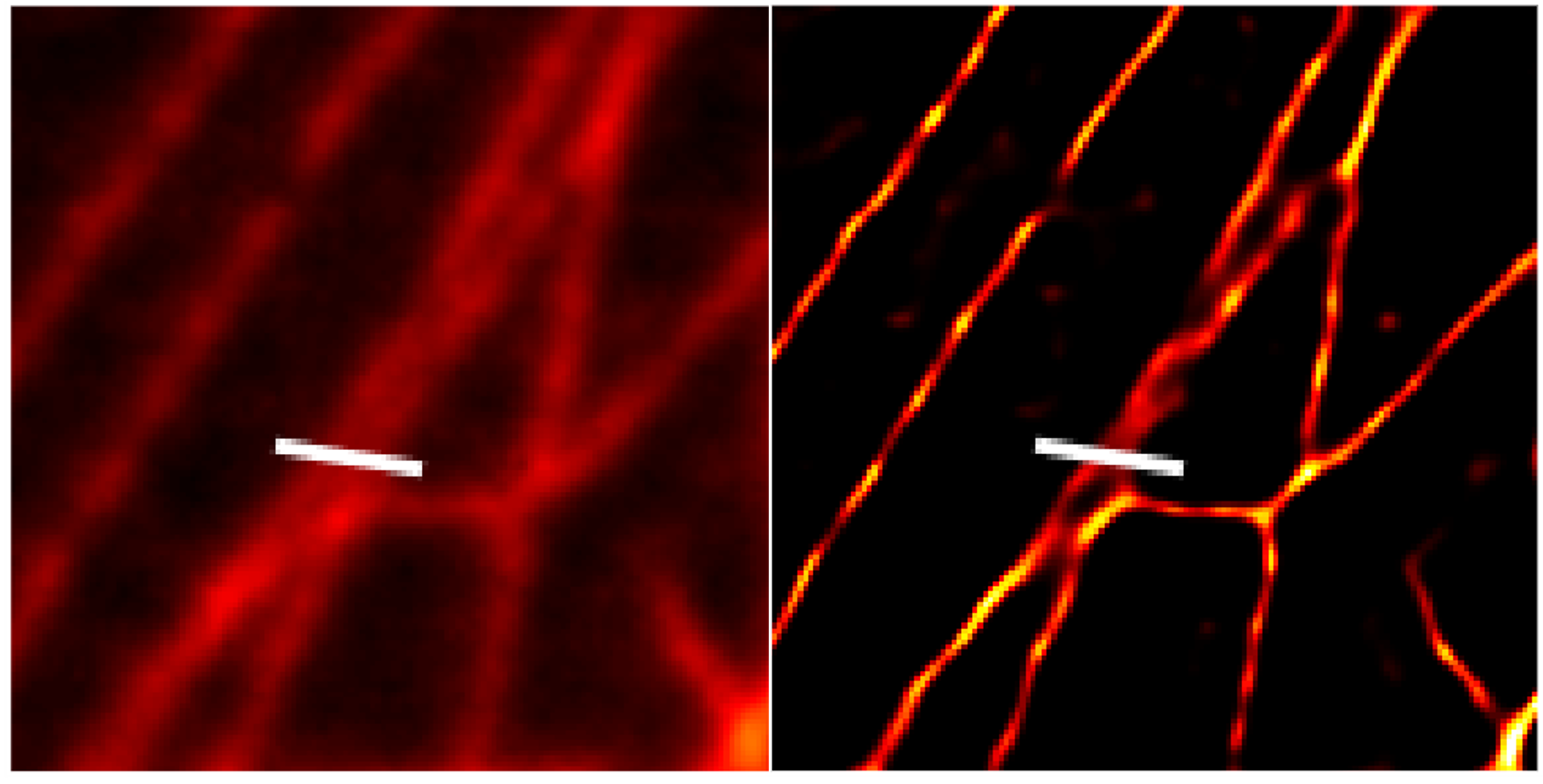
The Cell Xpanse system integrates SPIN SACD super-resolution spinning-disk technology, offering a combination of confocal imaging with two distinct super-resolution techniques.
SACD uses two-step deconvolution and autocorrelation to overcome diffraction limits, making it suitable for imaging thicker samples with resolution down to 120 nm.
MI-SIM enables further resolution improvements, supporting deeper insight into dynamic biological processes.
Having two super-resolution approaches allows researchers to cross-validate findings and increase confidence in results.
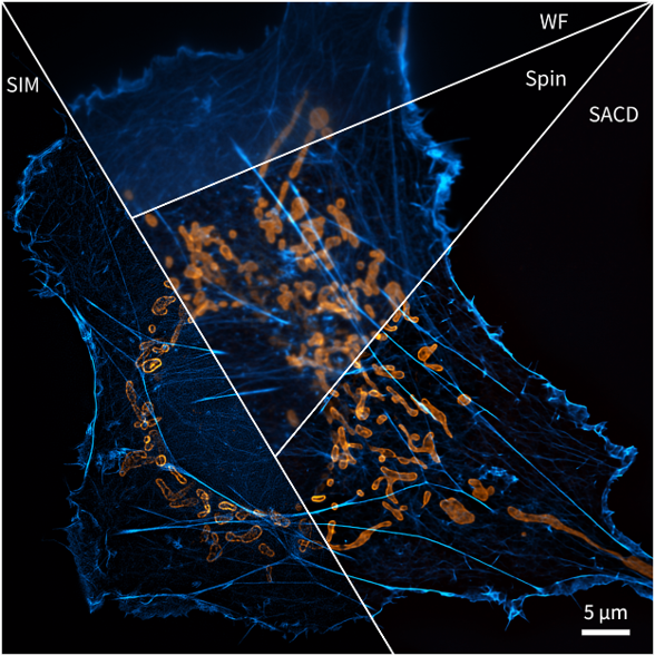
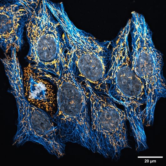
Figure shows a cell stained for actin, blue, and mitochondrial outer membrane, orange. Panels show the same cell in widefield, spinning-disk confocal, SPIN SACD and SIM, highlighting differences in resolution across imaging techniques.
Unparalleled versatility
The Cell Xpanse platform offers users unparalleled versatility, with one system able to image an enormous variety of biological sample types.
Due to the exceptional resolution, SIM is capable of imaging challenging small samples such as bacteria and yeast cells.
Furthermore, spinning disk offers an ideal high-throughput solution for imaging large-format 3D samples such as brain slices and model organisms.
Both techniques offer high temporal resolution (>100 fps) and low photostress, enabling the capture of highly dynamic events in live samples.
Due to the low photostress, both technologies are also ideal for long-duration time-lapse recordings.
Both SIM and spinning disk are camera-based techniques, offering a huge dynamic range. This enables quantitative image acquisition from complex biological samples containing multiple or dynamic protein expression levels.
Application images
Cell Xpanse is designed for researchers working across scale, from tissue architecture and model organisms to higher-resolution structural imaging within a single workflow.
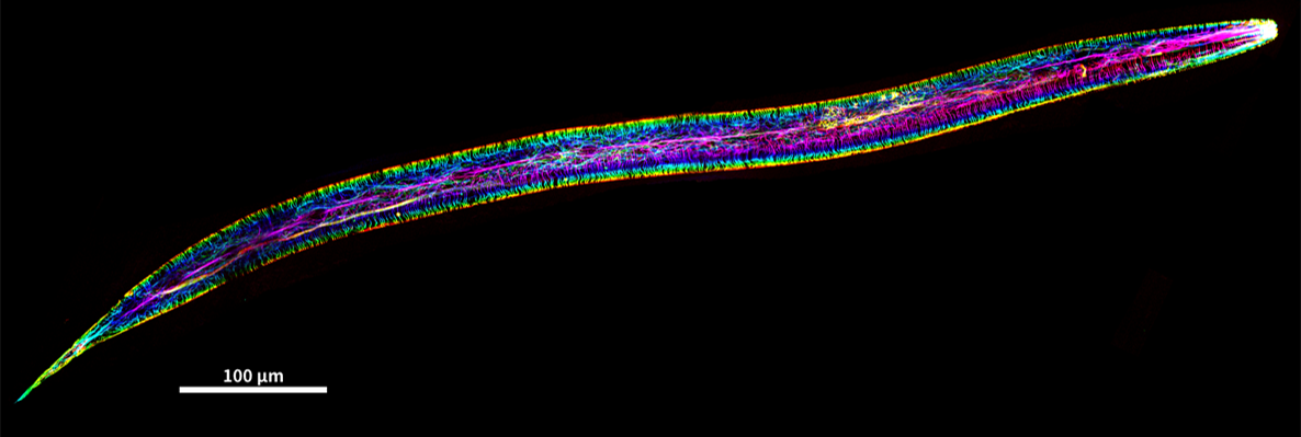
Spinning-disk image of C. elegans microtubule structure. source CSR Biotech
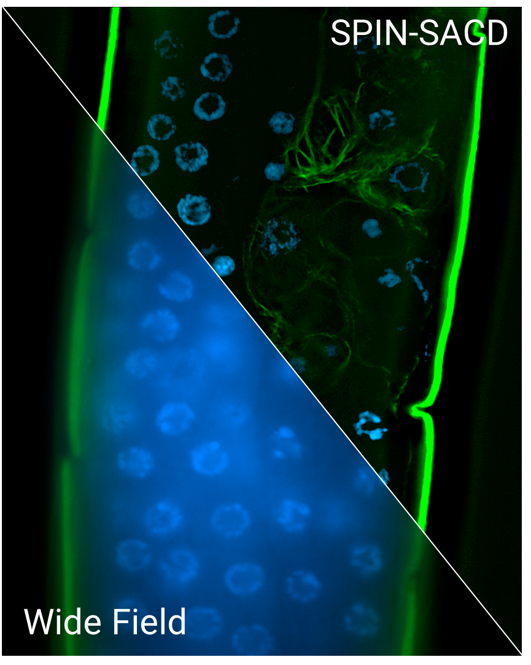
C. elegans SACD (DNA (blue) and actin (green). Source CSR Biotech
Expansion microscopy immune synapses between Jurkat T cell and Raji B cell labeled with pTyr (red), LAT (green) and the nucleus (blue). Source Dr. Delphine Judith, Institut Cochin, Paris
1000 µm Z-stack of Mouse heart blood vessels Source: Prof. Shen's group at Nanjing University
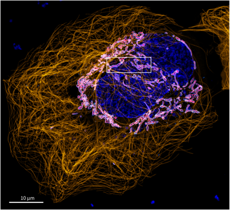
U2OS cells microtubule Orange, DAN (blue) Mitochondria Inner membrane(red) outer membrane (Cyan), SPIN Vs SPIN-SACD Source: CSR Biotech
Download the brochure
Download the Cell Xpanse brochure to learn more about our advanced cell analysis platform.