Technology overview

Imaging technologies for live-cell research

From fine fluorescent structures to label-free views of whole cells, CSR Biotech systems use complementary approaches to explore living samples. Discover the principles, imaging capabilities and processing methods behind each technology.

Explore the three imaging technologies

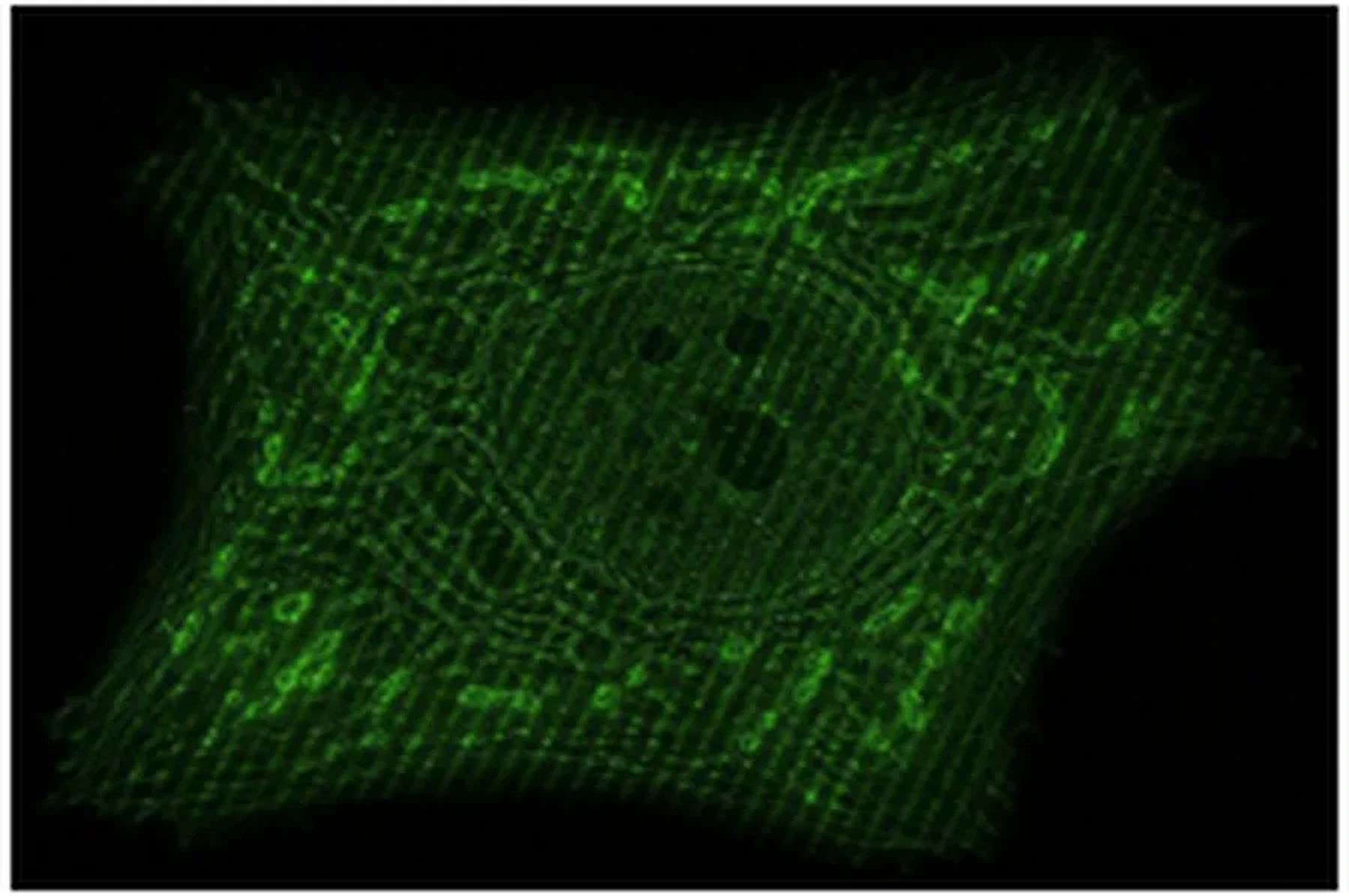
Green fluorescence image acquired using structured illumination microscopy

Structured illumination microscopy (SIM)

Live-cell super-resolution

SIM uses patterned illumination and computational reconstruction to reveal structures beyond the diffraction limit. Explore how illumination patterns, image acquisition and processing work together to enable super-resolution live-cell imaging.
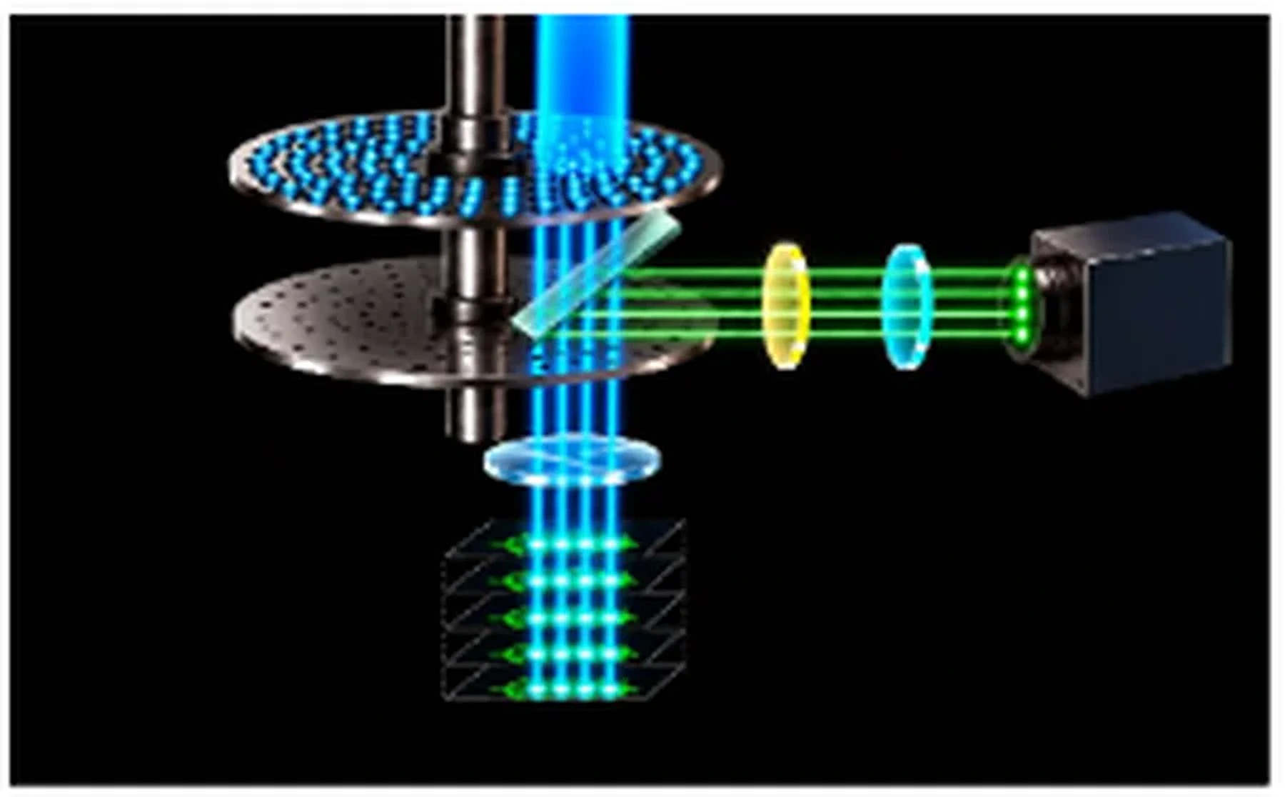
Optical diagram illustrating spinning-disk confocal microscopy

Spinning-disk confocal

High speed confocal

Spinning-disk confocal simultaneously scans multiple illumination points across the sample. The parallelized confocal scanning significancy increases through put and offers lower photo stress making ideal for live-cell imaging. Explore the impact of different disk designs and image processing techniques.

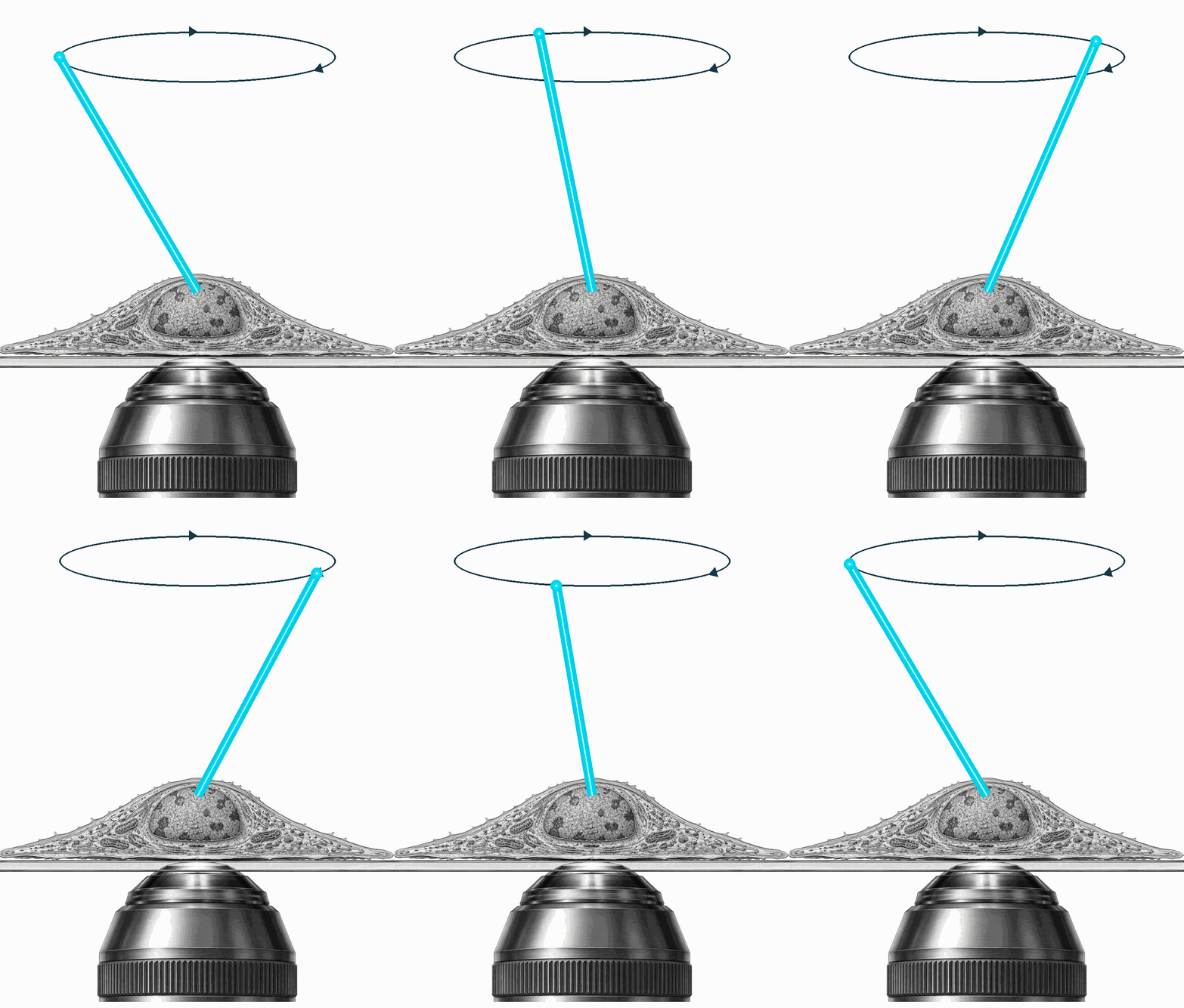
Optical diffraction tomography diagram showing illumination of a sample

Optical diffraction tomography (ODT)

Label-free super-resolution

ODT uses transmitted light to captures refractive-index differences in samples to reconstruct 3D images of un-labeled cell Explore how the technique works and how it complements fluorescence imaging in live-cell research.