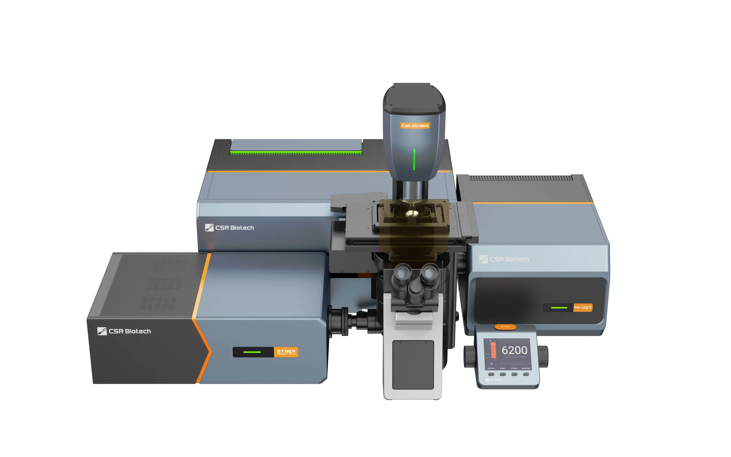
Key benefits
Correlative super-resolution
See the full picture by combining SIM with label-free imaging to give molecular dynamics greater spatial context.
Next generation live-cell imaging
HeLa Cell Mitosis, Technique: ODT, Source: CSR Biotech.
Next generation live-cell imaging workflows
Advanced AI-enabled software tools allow users to combine multiple imaging technologies in a single workflow for maximum impact.
Correlative super-resolution
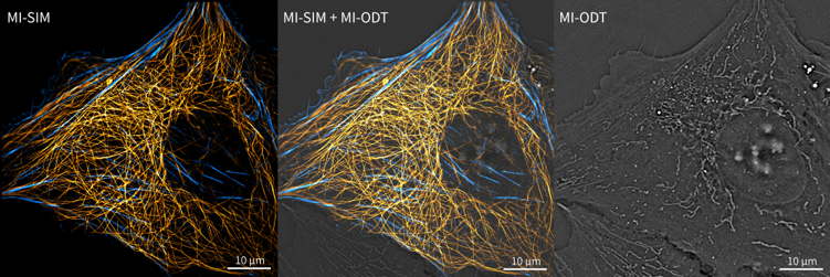
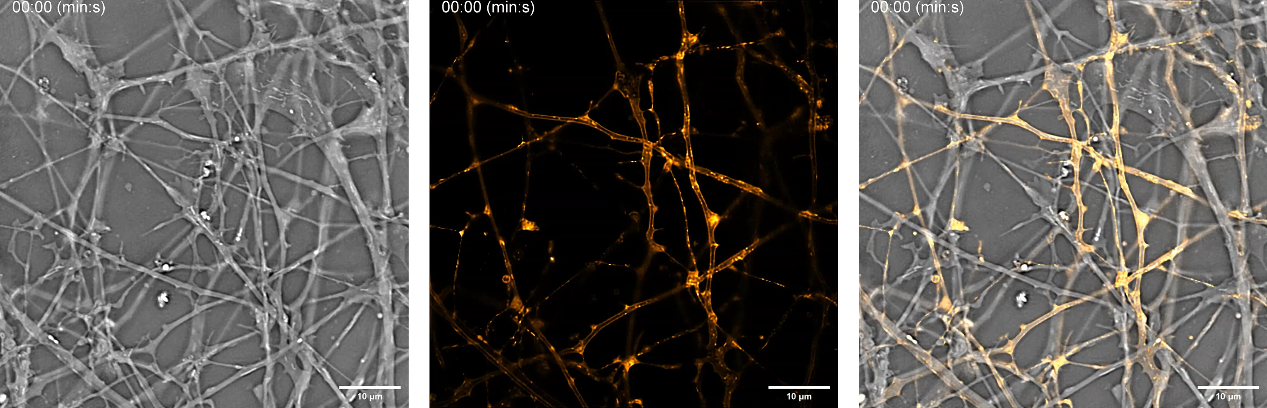
The integration of Optical Diffraction Tomography (ODT) with Structured Illumination Microscopy (SIM) creates a powerful synergy that advances cellular imaging.
- SIM enables users to localize and track a limited number of biomolecules specifically labeled with fluorescence dyes with extremely high spatial and temporal resolution.
- ODT enables users to visualize cellular morphology and the dynamics of unlabeled organelles based on the refractive index of the different cellular compartments, offering full spatial context.
- With Cell Xtreme, users gain a comprehensive, contextual view of cellular architecture combined with the pinpoint molecular specificity of SIM, unlocking deeper insights into biological processes.
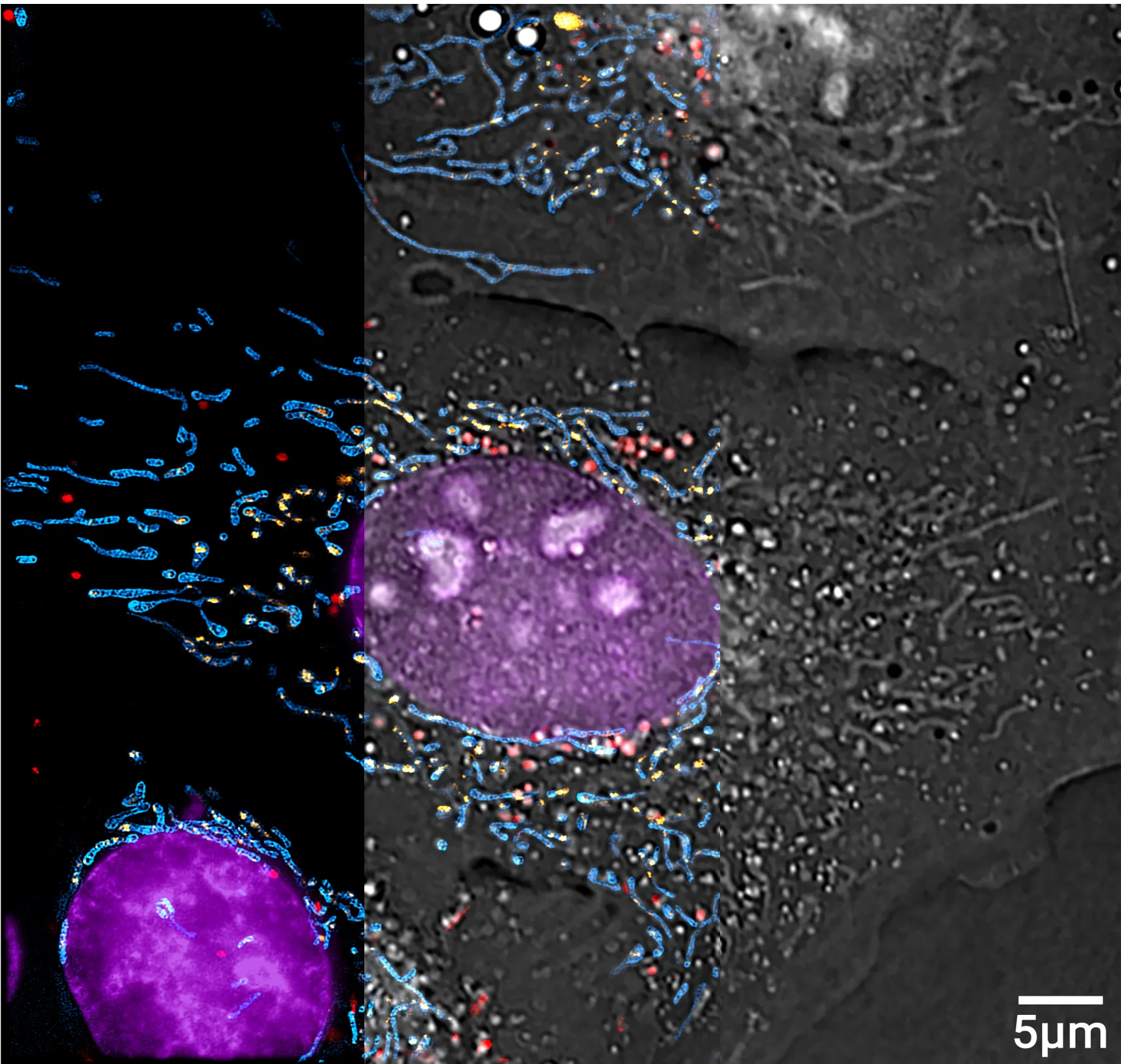
Figure shows U2OS cell stained for DNA, inner mitochondrial membrane, mitochondrial DNA and lysosomes, imaged with SIM and ODT. Merge panel shown in the center.
Next generation live-cell imaging
Label-free imaging offers a powerful advantage for modern microscopy by reducing dependency on fluorescent labels to visualize cellular structures.
- ODT spares researchers from the need to engineer fluorescent proteins or introduce potentially toxic dyes into their samples.
- By using transmitted light to reconstruct 3D refractive index maps of live samples, ODT removes the need for damaging high-intensity illumination inherent to fluorescent imaging.
- ODT causes minimal perturbation, preserving cell viability and enabling continuous long-term observations of live samples, as well as high-frequency observation of highly dynamic processes.
- ODT offers tomographic super-resolution, 130 nm, imaging technology able to acquire volumetric images in less than 1 second without the need for z-stacking.
- By relying on intrinsic optical properties of the sample, scientists gain a single, streamlined imaging technology capable of revealing multiple biological structures, including DNA, mitochondria, lipids and nucleoli, in detail.
AI segmentation
Imager software offers users trained AI segmentation tools capable of segmenting key cellular structures.
- Mitochondria
- The nucleus, chromatin
- Nucleoli
- Lipid droplets
- Cell boundaries
- When used in combination with SIM, users can use fluorescent imaging channels normally reserved for common cellular markers to label other biomolecules of interest, opening up the possibility of multiplexed live-cell imaging.
- Alternatively, users can reduce the number of fluorescent imaging channels required for an assay, reducing photo stress and extending the duration of their experiment.
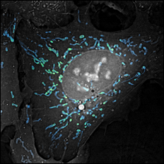
Figure shows AI segmentation of an ODT image, enabling users to generate masks of mitochondria, liposomes, nuclei, nucleoli and the cell boundary, removing the need to stain these organelles with fluorescent dyes.
Advanced system integration
Advanced system integration tools simplify multimodal imaging workflows and support efficient acquisition, alignment and analysis across imaging modes.
- Easy-to-use graphical multimodal imaging workflow setup.
- Digital image registration tools for simplified alignment and analysis.
- Multi-site and sample overview tools available for all imaging modes.
- Live-view ODT and SIM modes for sample positioning and monitoring.
- Fully integrated image processing and display tools for all imaging modes.
- AI-driven software enables customizable, multi-position monitoring and automated triggering of experiments, while seamlessly integrating and processing data from both imaging modalities.
Figure: Video shows real-time ODT live preview mode. Imager software allows users to adjust focus in real-time, scanning the top and bottom focal planes to identify targets.
Next generation live-cell imaging workflows
Feedback microscopy: Cell Xtreme enables highly dynamic imaging workflows by dynamically adapting imaging parameters in real-time. By utilizing intelligent software, the system adapts to the environment to capture rapid cellular events with reduced phototoxicity.
Multiplex live-cell imaging: Seamlessly observe multiple molecular targets at once. The combination of ODT and SIM empowers researchers to view unlabeled structures in context with specific fluorescent markers for comprehensive live-cell imaging.
Hybrid tracking capabilities: Confidently follow highly dynamic events over extended periods. Our approach allows for high-frequency tracking using low-phototoxicity techniques while integrating targeted, high-resolution snapshots to balance image resolution with cell viability.
Figure shows a hybrid tracking mode, AI segmentation of an ODT image enables users to track mitochondria dynamics with high temporal resolution at 10 second intervals. Every 4th image a high-resolution SIM image is acquired enabling users to balance resolution and photo stress.
Application gallery
Explore results showcasing the capabilities of our systems across real-world applications.
Download the brochure
Get detailed information about Cell Xtreme capabilities and features.