MI–SIM

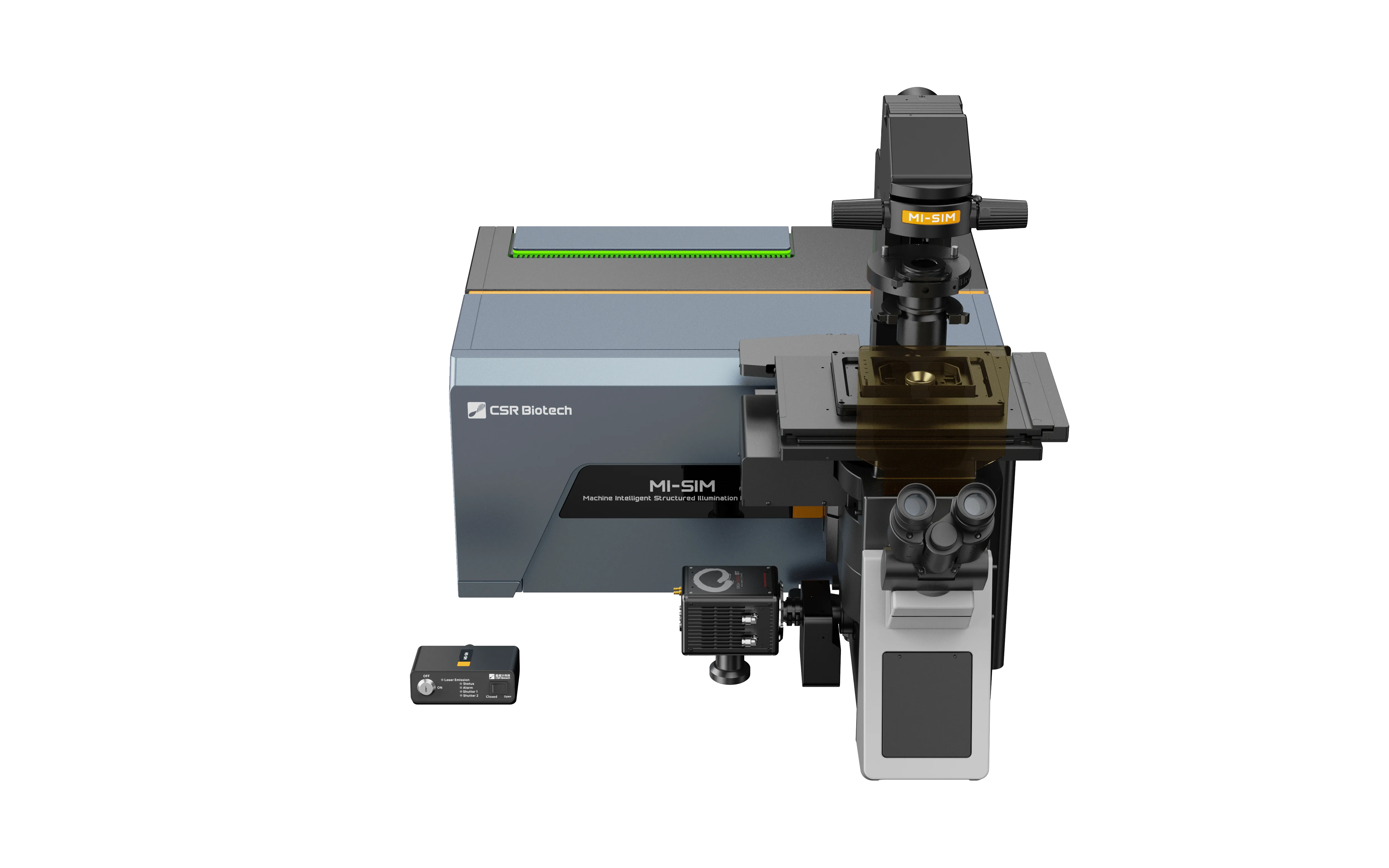
MI–SIM offers a next–generation structured illumination microscope designed for live-cell research, combining extremely high spatial and temporal resolution with low photo-stress imaging in a flexible, user-friendly platform.

MI–SIM

Key benefits

Next–generation SIM resolution

Next–generation SIM resolution

With data showing 60 nm resolution in X–Y and 200 nm in Z, MI–SIM enables researchers to resolve fine subcellular structures beyond the reach of diffraction-limited microscopy systems.

Designed for live samples

2D-SIM imaging in live U2OS cells reveals dynamic interactions between mitochondria (cyan) and the endoplasmic reticulum (yellow), enabling rapid visualization of organelle behavior and direct observation of contact sites between different organelles.

Multiple imaging modes

2D, 3D and TIRF SIM options give researchers flexibility across different imaging workflows.

Resolving power

Next–generation resolving power

MI–SIM offers extremely high resolution, achieving 60 nm resolution in X-Y and 200 nm in Z, significantly improving on the performance of legacy SIM platforms.

The system uses a spatial light modulator to illuminate the sample with a high-frequency SIM pattern, then applies advanced processing algorithms including Hessian SIM and SPARSE deconvolution to extend resolution performance further.

This enables users to resolve fine subcellular structures including mitochondrial cristae, endoplasmic reticulum, and clathrin-coated pits in live dynamic cells, supporting deeper insight into biological processes.

Endoplasmic reticulum in live U2OS cell, CSR Biotech

Live-cell imaging

Live-cell compatible super-resolution

SIM technology offers a highly sensitive imaging approach compatible with conventional live-cell dyes and fluorescent proteins, making it well suited to live-cell imaging.

Unlike traditional confocal systems that rely on pinholes to block signal, SIM is a widefield technique that captures a greater proportion of emitted photons. This allows SIM to operate at lower excitation powers, helping reduce bleaching and phototoxicity.

The rapid pattern switching of the spatial light modulator, combined with modern high-performance sCMOS cameras, enables extremely high temporal resolution of over 300 reconstructed frames per second. As a result, researchers can capture fast subcellular dynamics over extended periods below the diffraction limit.

Multi-color time-lapse imaging of DRP1 foci, which play a critical role in mitochondrial dynamics, the outer mitochondrial membrane, and the endoplasmic reticulum in live U2OS cells. Imaging was performed under conditions that minimize photobleaching and phototoxicity, allowing long-term observation of organelle dynamics in multiple colors. CSR Biotech

Imaging modes

Multimodal SIM

MI–SIM provides a range of SIM illumination modes, allowing users to tailor acquisition to specific experimental needs.

  • 2D SIM is optimized for high temporal resolution.
  • 3D SIM is optimized for improved Z resolution.
  • TIRF SIM is optimized for signal-to-noise performance near the coverslip.
  • This flexibility helps users generate the right data from different cellular compartments and experimental set-ups.
1 of 3

TIRF SIM

Imaging of microfilaments in live RBL–2H3 cells with TIRF–SIM modality, enabling high signal-to-noise visualization of microfilament dynamics, particularly for membrane-associated events occurring near the coverslip. Dr Kseniya Korobchevskaya, KIR platform, University of Oxford

Dual camera module

SYNER dual camera module

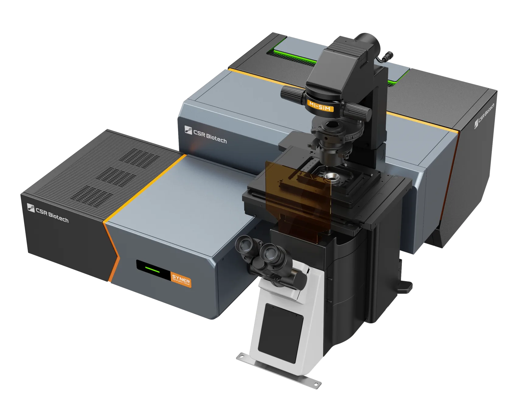
MI–SIM offers the option to include the SYNER dual camera module, supporting high-speed sequential or simultaneous dual-channel SIM acquisitions.

SYNER includes simple digital image alignment tools, helping maintain consistency across channels.

Unlike many dual camera systems, SYNER does not limit workflows to two fixed channels. With dual filter wheels and motorized dichroic switching, users can move between single-camera bypass mode and high-speed multi-channel acquisitions with more than four channels.

SYNER dual camera module
Software

Imager and FINER software

  • MI–SIM includes intuitive acquisition and processing software through Imager and FINER.
  • Sample overview functions and real-time super-resolution help users identify areas of interest quickly.
  • Expandable functionality supports advanced workflows including multi-position, multimodal and event-driven multi-phase experiments.
  • Integrated image processing tools support acquisition, reconstruction and analysis workflows.
  • Intelligent features include cell tracking, AI-based object recognition and event-based triggering for feedback microscopy.
  • Imager and FINER output standard TIF images compatible with third-party analysis software.

Overnight time-lapse imaging of bone marrow stem cell migration across multiple sites, enabling high-throughput, simultaneous tracking of cellular dynamics. Zhang Lab, Xi'an Medical University

Extended gallery

MI–SIM gallery

Explore a wide range of applications and sample types captured using the MI–SIM platform.

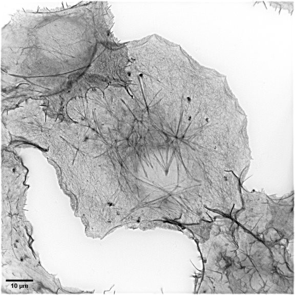
2D TIRF-SIM enables high-resolution imaging of actin cytoskeleton (black), clearly delineating stress fibers and intricate substructures to provide deeper insight into cellular architecture at the membrane.

2D TIRF-SIM enables high-resolution imaging of actin cytoskeleton (black), clearly delineating stress fibers and intricate substructures to provide deeper insight into cellular architecture at the membrane.

TIRF-SIM COS-7 Cells Actin labeled with, SPY488-FastAct (Spirochrome), CSR Biotech
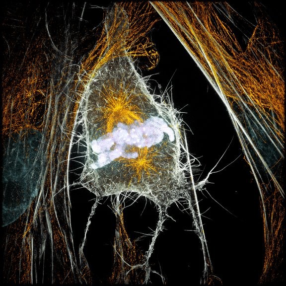
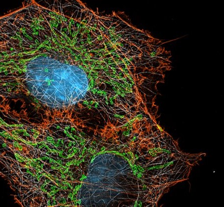
High-resolution 2D-SIM image achieved only after Wiener reconstruction, highlighting mitosis of Indian muntjac fibroblasts with microtubules (yellow), chromosome (violet), kinetochores (white) and actin microfilaments/stress fibers (gray).

High-resolution 2D-SIM image achieved only after Wiener reconstruction, highlighting mitosis of Indian muntjac fibroblasts with microtubules (yellow), chromosome (violet), kinetochores (white) and actin microfilaments/stress fibers (gray).

2D-SIM of Indian muntjac fibroblasts microtubules (yellow), chromosome (violet), kinetochores (white) and actin (gray). Dr. Helder Maiato, i3S, University of Porto, Portugal

2D-SIM mitochondrial DNA Blue neuronal membrane (yellow)

Shenzhen Institutes of Advanced Technology (SIAT), Prof. Dr. Qiao Zhao

3D-SIM mitochondrial inner membrane (IMM, green) outer membrane (OMM, red)  mitochondrial DNA (mtDNA, cyan).

3D-SIM mitochondrial inner membrane (IMM, green) outer membrane (OMM, red) mitochondrial DNA (mtDNA, cyan).

Song Lab, Wuhan University

2D-SIM Cyanobacteria, cytosolic label (red) and Glucose pathway-specific label (green)

Zhao Lab, Shandong Normal University

3D-SIM imaging reveals the periodic arrangement of spectrin and actin, providing high-resolution insight into the nanoscale organization of the neuronal cytoskeleton.

3D-SIM imaging reveals the periodic arrangement of spectrin and actin, providing high-resolution insight into the nanoscale organization of the neuronal cytoskeleton.

Spectrin (blue) and Actin (red) in fixed neurons from rat. Dr. Ana Rita Costa, i3S - Nerve Regeneration Group, University of Porto

Download the brochure

Receive more information on MI–SIM, request a quote, or speak with our team about the right workflow for your samples.

Ready to see MI–SIM in action?

Book a demo to experience the technology firsthand, or get in touch with our team to discuss your research needs.