Optical diffraction tomography (ODT)
Optical Diffraction Tomography (ODT) generates volumetric, label-free images using transmitted light to capture refractive-index variations in different cellular compartments. Computational image processing in Fourier space converts this information into super-resolved images. ODT data can be segmented with AI to visualize discrete cellular compartments, including the nucleus, liposomes and mitochondria. ODT can also be combined with fluorescent techniques such as spinning disk and SIM to provide additional spatial context for live-cell data without subjecting samples to significant additional photostress.
Why ODT is ideal for live-cell imaging
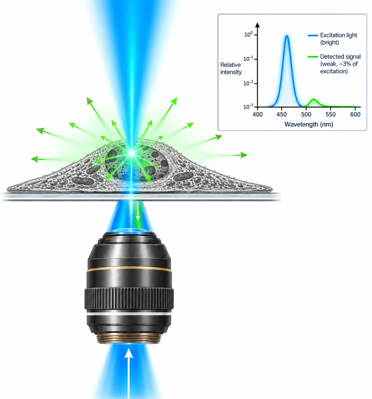
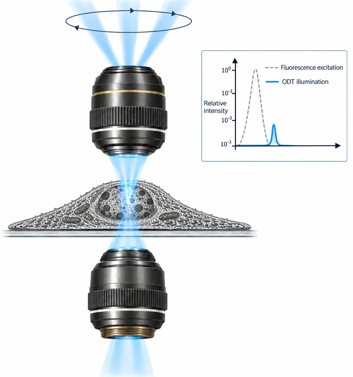
Optical Diffraction Tomography (ODT) generates images using transmitted light rather than detecting emissions from fluorescent dyes. With transmitted-light techniques, more than 90% of the light used to illuminate the sample is captured to form the image. With fluorescence microscopy, only a small percentage of the excitation light is absorbed by a fluorescent dye, and only a small proportion of the emitted fluorescent signal is detected. Fluorescence imaging therefore requires high illumination intensities to generate sufficient signal, leading to phototoxicity and photobleaching in live specimens. Because ODT requires low illumination power, it is more light-efficient and reduces photostress, making it well suited to live-cell imaging.
ODT: how it works
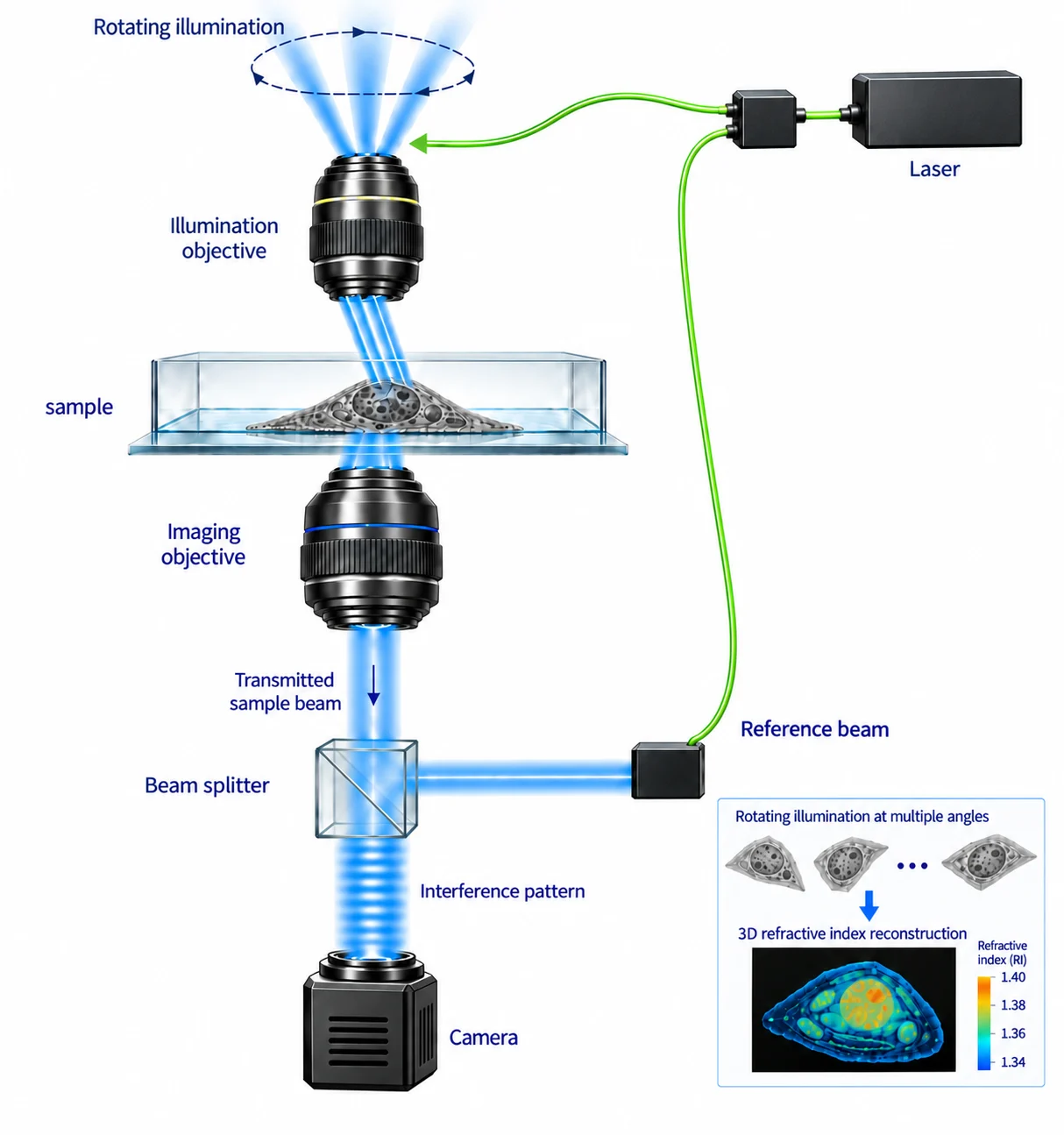
ODT is a tomographic imaging technique similar to CT scanning, but instead of using X-rays it uses light. ODT employs a coherent light source, such as a laser, split into two optical paths. One beam illuminates the sample while the second reference beam travels through an interferometric arm. The sample is illuminated from multiple angles using a high-numerical-aperture objective positioned above the sample. The beam passes through the sample and is collected by a second objective. The imaging and reference beams are recombined and recorded as holographic interference patterns by a high-speed camera. The amplitude and phase shifts introduced to the imaging beam by variations in the sample’s refractive index are then computationally processed into the ODT image in Fourier space. The grayscale values in the resulting ODT image indicate relative refractive-index variations.
Cell Xtreme acquisition modes
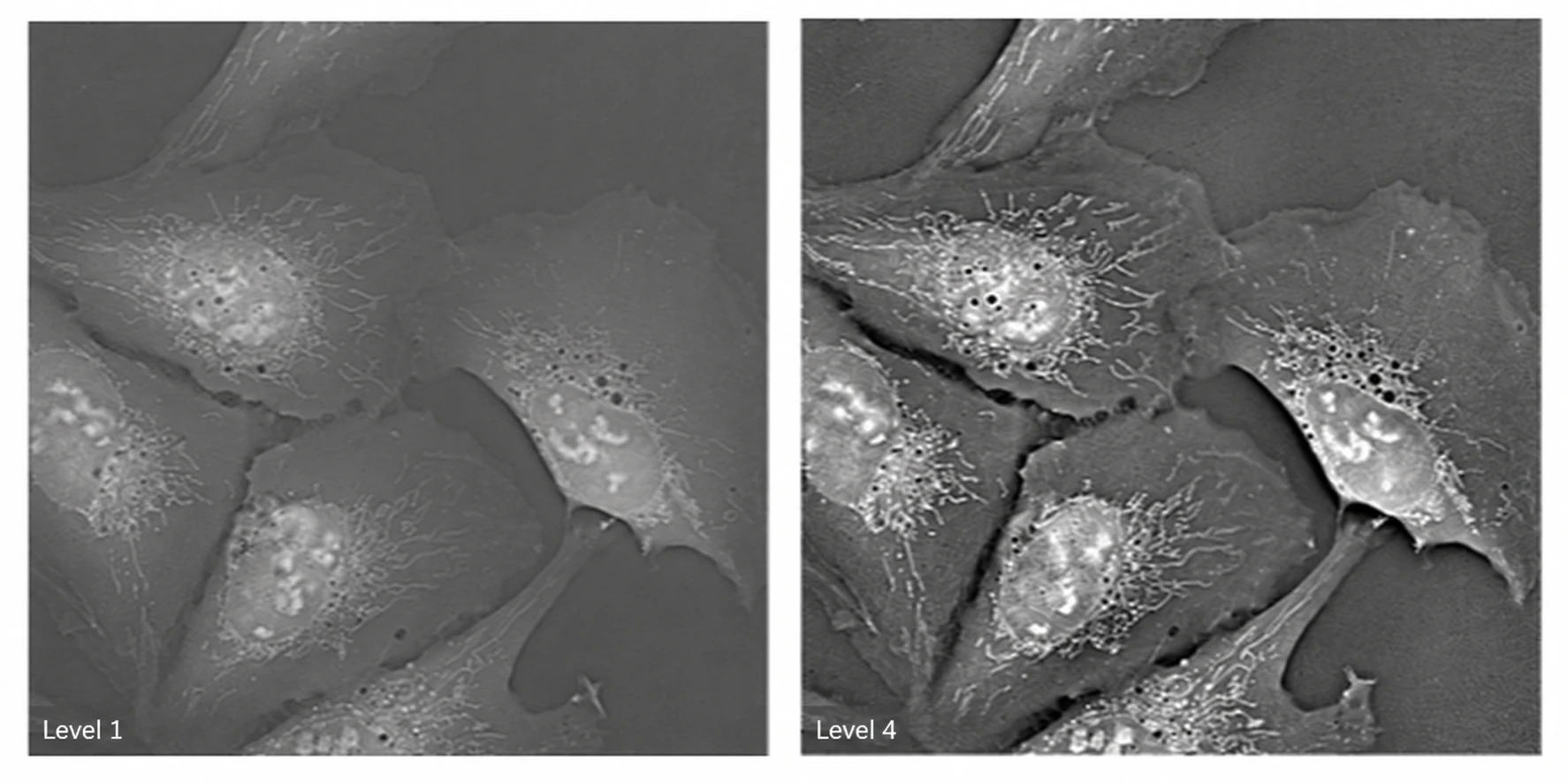
The Cell Xtreme system offers multiple acquisition modes, allowing users to choose between speed and resolution. Level 1 prioritizes speed, capturing images at 60 angles per 360-degree scan to shorten acquisition and processing time.
At Level 4, the system captures 360 separate images in approximately one second. The additional information improves image resolution and contrast, offering lateral resolution down to 130 nm.
Applications in biological and biomedical imaging
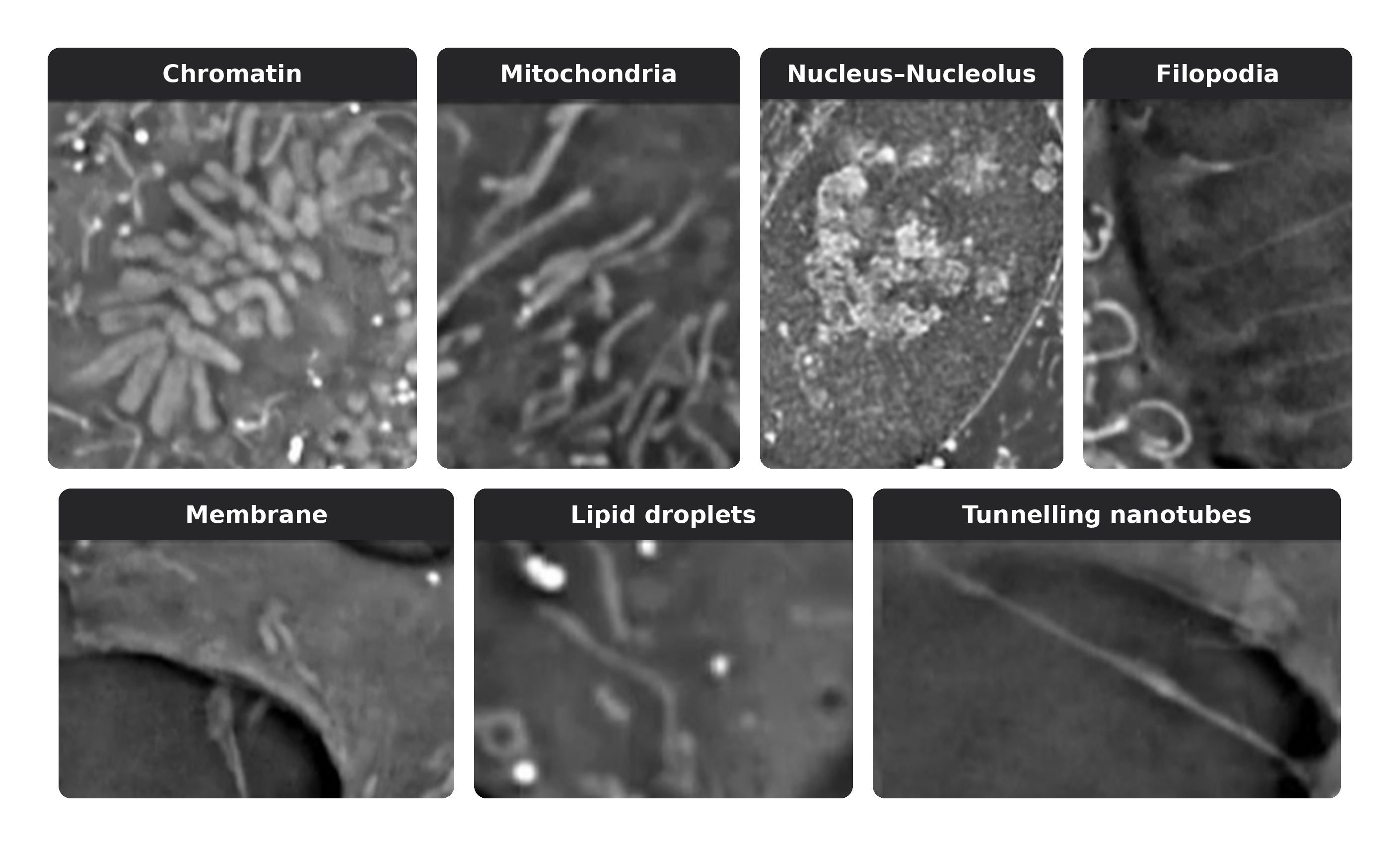
ODT’s combination of high spatial resolution, label-free contrast, and gentle illumination makes it a powerful tool in life sciences. It enables quantitative visualization of intracellular organelle organization and dynamic processes such as mitosis and organelle dynamics without the need for fluorescent labels. By capturing refractive index variations, ODT can differentiate subcellular structures, track morphological changes over time.
AI image segmentation
AI models can be used to segment Specific cellular compartments based on morphology and refractive index differences. In these images the cell boundary, lipid droplets, mitochondria the nucleus and nucleoli have been segmented. AI segmented masks means researchers no longer need a separate fluorescent markers to identify these organelles’ enabling them to use fewer fluorescent channels reducing photo stress. Once assays have been validated, in some cases experiments can be performed solely using ODT.
ODT hybrid imaging offers the best of both worlds
ODT can be combined with fluorescence techniques such as SIM or spinning-disk confocal microscopy in a single hybrid imaging workflow. Fluorescence enables the tracking and monitoring of interactions between tagged molecules. ODT provides spatial context and additional information about the location, morphology and dynamics of other subcellular compartments.
In some cases, AI masks enable ODT to replace a fluorescence channel, reducing photostress. Combined with AI-based event detection, ODT can support advanced multiphase workflows.