Structured illumination microscopy
Structured illumination microscopy (SIM)
SIM is a powerful super-resolution technique that uses patterned illumination and computational image processing to recover detail beyond the diffraction limit. It is well suited to live-cell work, combining improved resolution with gentle, dynamic imaging.
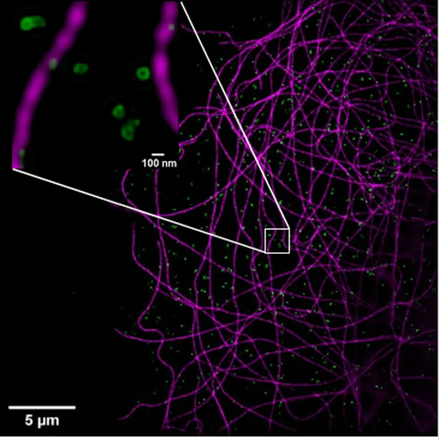
Structured illumination microscopy demonstration
How to use moiré fringes to break the diffraction limit
A moiré fringe pattern that occurs when two high frequency patterns are overlaid. In SIM, moiré fringes arise from the interaction of the illumination pattern with high resolution structures in the sample. As the moiré fringe patterns are lower resolution than the structures that create them, this otherwise unresolvable information can now be transmitted through diffraction limited optics.
Phase shifting and rotating the illumination pattern gives access to information from structure in the sample with different phases and orientations.
As we know the Illumination pattern that creates the moiré fringe, by analyzing the additional information in Fourier space, we can computationally reconstruct fine structural details in the sample that lie below the diffraction limit.
Introduction to Fourier space
SIM image reconstruction occurs in Fourier space, where images are represented in terms of spatial frequencies rather than spatial positions. Each point in Fourier space corresponds to a specific spatial frequency and orientation.
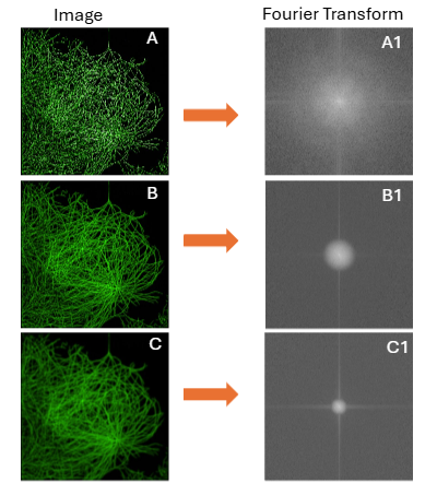
The further the away from the center of the Fourier spectrum, the information is located, the higher the spatial frequencies/ resolution of the information. Image A represents an original image with unlimited resolution and its corresponding Fourier transform.
If the same image is captured by an optical system, the numerical aperture of the objective limits the resolution of the resulting image constraining the Fourier transform (image B-B1).
If an objective has a lower NA the resulting image will have lower resolution and the Fourier transform will be further constrained (Image C-C1).
SIM image acquisition
SIM image acquisition
Rotating and phase-shifting the illumination pattern generates multiple orientations and phases. Because resolution enhancement occurs only perpendicular to the stripe direction, at least three orientations separated by 60° are required for isotropic lateral resolution improvement. Depending on the imaging mode, a complete SIM reconstruction requires 9 raw images for 2D SIM or 15 for 3D SIM.
SIM illumination control
In MI-SIM, the sinusoidal illumination pattern is generated by projecting a laser onto a spatial light modulator (SLM), which introduces a diffraction pattern into the beam. The light is split into two separate beams and focused into the back aperture of the objective; the beams interfere in the sample plane to form a sinusoidal striped illumination pattern.
By adjusting the beam positions in the back focal plane, the system can operate in epi-illumination, highly inclined illumination (Hi-Lo), or total internal reflection fluorescence (TIRF) modes, enabling a trade-off between sample penetration depth and signal-to-noise ratio.
Incorporating a third interfering beam enables 3D SIM by producing a volumetric interference pattern, improving both axial resolution and optical sectioning.
Comparison of 2D–SIM, TIRF–SIM and 3D–SIM
2D–SIM
TIRF–SIM
3D–SIM
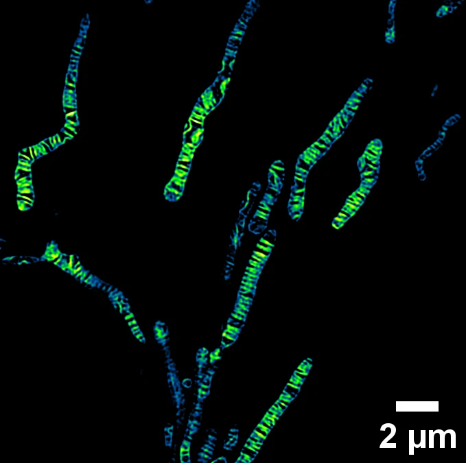
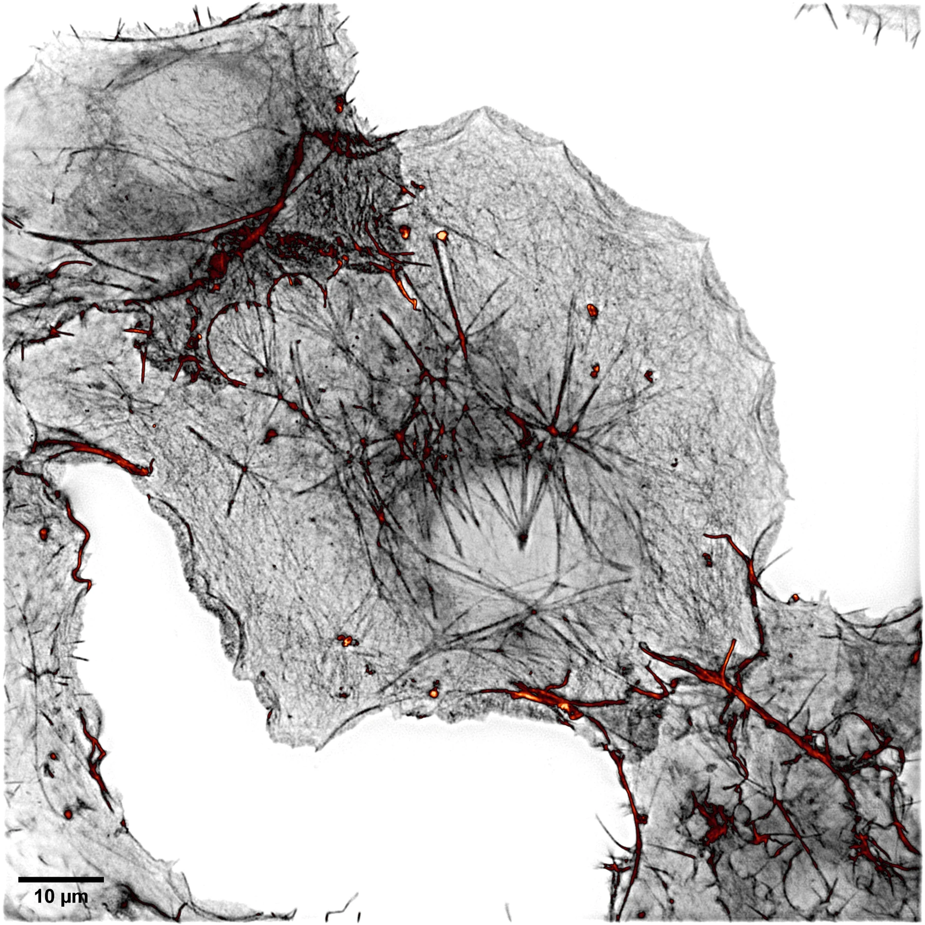
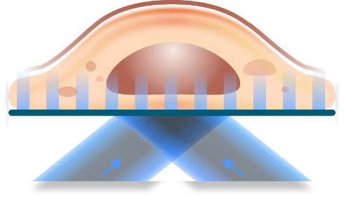
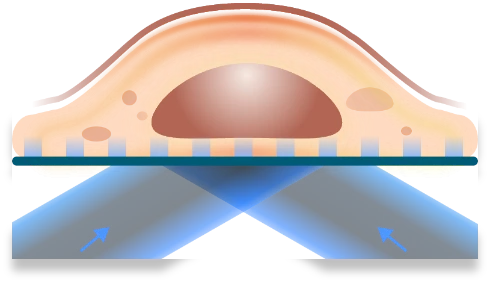
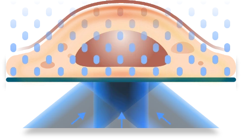
SIM acquisition and Fourier space
By acquiring multiple phase-shifted images for each illumination orientation, the full Moiré information is sampled, allowing the shifted frequency components to be separated and repositioned to their correct locations in Fourier space. Using at least three stripe orientations provides nearly complete circular coverage of Fourier space. The fundamental resolution limit of SIM is set by the requirement that the illumination pattern frequency itself must lie within the resolution of the diffraction-limited optical system. With the additional information contained within the Moiré fringes, SIM can achieve up to a twofold improvement in spatial resolution compared to conventional widefield microscopy.
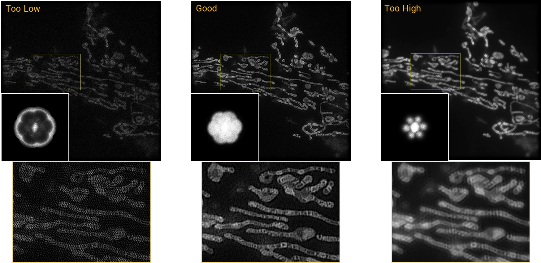
Image reconstruction and filtering
The raw SIM images are combined into a single composite spectrum in Fourier space. During this process, higher spatial frequencies are often disproportionately amplified, which can increase noise in the reconstructed image. To rebalance the spectral components, a deconvolution step is applied using a Wiener filter. When set too low, high-frequency noise dominates the reconstruction; when set too high, genuine high-frequency information is suppressed, resulting in reduced resolution.
Pushing the resolution further with sparse deconvolution
Sparse deconvolution factors in the continuity of biological structures and the sparsity of the distribution of excited fluorophores. By applying a Sparse deconvolution to the reconstructed SIM data, MI-SIM can further improve the lateral resolutions (~60 nm) and boost image contrast.
fluorescence microscopy. Nature Biotechnology 40, 606–617 (2021).